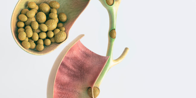
 The three essential components of human diet are protein, carbohydrate, and fat, along with vitamins and minerals. To digest and absorb most of the dietary fat from the intestine, bile is needed. Bile is constantly formed by the liver. Bile thus formed is normally carried away from the liver by the bile canaliculi, which join together to form the right and left hepatic ducts within the liver. These ducts, after exiting the liver hilum, join together to form a common hepatic duct that traverses down to the small intestine (duodenum), where it opens up to pour the bile as and when needed to digest and absorb the consumed fat. Normally, bile from the liver does not go straight down to the intestine, but gets stored and concentrated in a pear-shaped leathery kind of bag attached to the under surface of the liver and connected to the common bile duct via a tube called cystic duct.
 Gallstone disease denotes illness caused by the presence of gallstone, either in the gallbladder or the bile duct. A gallstone is a stone formed within the gallbladder out of bile components, which are bilirubin pigment, cholesterol, and bile salts. Gallstones formed mainly from cholesterol are termed cholesterol stones, and those mainly from bilirubin are termed pigment stones.
Most people with gallstones, almost 80-90 percent, never develop symptoms or illness, as such. They are just there, ending up in the grave or cremation after death! Illness typically starts when a stone blocks partially or completely the flow of bile down to the intestine. Blockage to the flow of bile may occur either at the gall bladder neck or the cystic duct before its entry into the bile duct, or at the level of the common bile duct. Occasionally, stones form in the portion of the biliary tree within the liver, and symptoms may develop if the stone blocks the flow of bile at the level of the common hepatic duct, or even higher up.
Main symptom of gallstone disease is a gradually building-up of severe cramp-like pain that starts in the midline, just below the right ribcage. The pain usually lasts for a few hours before subsiding gradually. Relief of pain is associated with the stone falling back to the gallbladder cavity, which unblocks the bile flow. This pattern is termed biliary colic, and usually lasts for less than six hours.
Alternatively, pain may shift or spread to the right upper part of the abdomen and remain persistent, making that part of the abdomen rigid and sore to touch or to mild pressure. This kind of illness is termed acute cholecystitis. Body temperature may rise gradually to above 100.4° F (38° C) in about one third of people with acute cholecystitis. The fever may be accompanied by chills. Acute cholecystitis happens if the block becomes established to set in inflammation of the inner lining of the gallbladder.
Similarly, if the stone blocks the bile duct, its lining gets inflamed, with no bile going to the intestine. Backflow of the bile pigment, bilirubin, into the blood stream leads to the yellow discoloration of the eyes, skin, and urine. Stool loses its normal light-yellow hue and becomes clay-like pale. This kind of presentation of gallstone disease is termed acute cholangitis.
Symptoms develop in about 1–4% of those with gallstones each year.
Chronic cholecystitis means persistently inflamed gallbladder. In this condition, the gallbladder is damaged by repeated attacks of acute inflammation, resulting over time in the bladder becoming thick-walled, scarred, and small. Cystic duct, or its opening, may get permanently blocked by the stone(s). The gallbladder may also contain sludge or stones. After prolonged period of inflammation, calcium may get deposited in the walls of the gallbladder, causing them to harden (called porcelain gallbladder). Porcelain gallbladder has a potential for the development of gallbladder cancer, in future.
If the bile duct gets blocked by the stone at or near its opening into the small intestine, it may also interfere with the flow of the pancreatic juice to the small intestine from the pancreas, the elongated gland that resides just below the stomach. If this happens, the backflow of the juice into the pancreas can set in acute inflammation of the pancreas itself. This condition is termed acute biliary pancreatitis, and may be lethal if not treated early and effectively.
Delayed diagnosis and treatment of acute cholecystitis can occasionally lead to the accumulation of pus in the gallbladder (empyema of the gallbladder). Pus collection under tension can lead to perforation of the gallbladder. These are serious life-threatening septic conditions.
Diagnosis of gallstone disease has been made easy by the advent and regular use of ultrasound scanning of the abdomen. Stones in the gallbladder are very rarely missed by a good ultrasound operator. Abdominal ultrasound examination can, however, miss stone in the bile duct in some fifty percent of cases. In such cases, special blood biochemistry tests give a lead to the evaluating physician, who can then request more specialized imaging study, such as MRI or an ultrasound examination from within the stomach or small intestine with a probe attached to an endoscope. This procedure is called endoscopic ultrasound scanning (EUS) or endosonography.
 Treatment
Biliary colic can be diagnosed and managed successfully in the emergency room (ER) of any general hospital. Blood tests and a few hours of observation will rule out acute cholecystitis, cholangitis, or pancreatitis. Patient can be discharged from the ER with a convenient date for surgery to remove the gallbladder. Patient should be counselled to restrict fat in diet until surgery.
Acute cholecystitis, or cholangitis cases need to be hospitalized for giving painkillers, antibiotics, fluid, and electrolytes by intravenous route, and to give rest to the bowel and biliary tract by no food intake by mouth for two to three days.
Acute cholecystitis usually resolves with this treatment alone, but this would be the right opportunity for early removal of the gallbladder, as well to prevent another attack. If patients with acute cholecystitis present a little late, surgery is deferred to six to eight weeks after the acute attack subsides completely.
Acute cholangitis may not resolve with intravenous treatment with antibiotics, fluids, and electrolytes alone. The stone blocking the bile duct needs to come out of the bile duct and the bile flow into the intestine needs to be re-established. This is done by a special endoscopic surgery called ERC (endoscopic retrograde cholangiography) and stone extraction into the small intestine. Endoscope is guided into the area of the small intestine where the bile duct joins the intestine. Through the endoscope, a deflated balloon is guided into the bile duct to the point above the blockage, and then the inflated balloon is carefully drawn back to the intestine. This procedure will pull the blocking stone down to the intestine and re-establish the flow of bile. After this procedure and resolution of cholangitis, patient should be advised surgery for the removal of the gallbladder to prevent further attacks.
 Medicosnext


