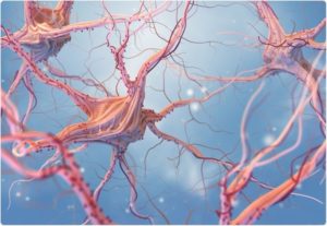
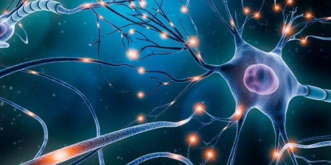
 The human brain is a complex organ that controls thought, memory, emotion, touch, motor skills, vision, breathing, temperature, hunger, and every process that regulates our body. It roughly weighs about three pounds in the average adult and is mostly composed of fat. It contains an estimated 86 billion neurons. Those billion of cells communicate by passing chemical messages at the synapse in a process called neurotransmission. This regular, rapid-fire messaging plays a big role in how you feel and function each day. By definition, synapse (also called as neuronal junction) is the site of transmission of electric nerve impulses between two nerve cells (neurons), or between a neuron and a gland or muscle cell (effector). A synaptic connection between a neuron and a muscle cell is called a neuromuscular junction.
At a chemical synapse, each ending, or terminal, of a nerve fiber (presynaptic fiber) swells to form a knoblike structure that is separated from the fiber of an adjacent neuron, called a postsynaptic fiber, by a microscopic space called the synaptic cleft. The typical synaptic cleft is about 0.02 micron wide. The arrival of a nerve impulse at the presynaptic membrane of membrane-bound sacs, or synaptic vesicles, fuse with the membrane and release a chemical substance called a neurotransmitter. This substance transmits the nerve impulse to the postsynaptic fiber by diffusing across the synaptic cleft and binding to receptor molecules on the postsynaptic membrane.
Neurotransmitters are chemical messengers that transmit as signal from a neuron across the synapse to a target cell, which may be another neuron, a muscle cell, or a gland cell. Neurotransmitters are chemical substances made by the neuron specifically to transmit a message. These are essential to the function of complex neural systems. A neurotransmitter can influence the function of a neuron through a remarkable number of mechanisms. A neurotransmitter acts in only of two ways: excitatory or inhibitory. These powerful neurochemicals are at the center of neurotransmission, and, as such, are critical to human cognition and behavior. A neurotransmitter influences trans-membrane ion flow either to increase (excitatory) or to decrease (inhibitory) the probability that the cell with which it comes in contact will produce an action potential. Type I synapses are excitatory in their actions, whereas type II synapses are inhibitory. Each type has a different appearance and is located on different parts of the neurons under its influence. The different types of neurotransmitters are discussed below.
Acetylcholine
Acetylcholine (Ach) was the first neurotransmitter discovered. It is a direct action small-molecule that works primarily in muscles, helping to translate our intentions to move into actual actions as signals are passed from the neurons into the muscle fiber. It also has other roles in the brain, including helping direct attention and playing a key role in facilitating neuroplasticity across the cortex.
Dopamine
Dopamine is often referred to as the “pleasure chemical”, because it is released when mammals receive a reward in response to their behavior; that reward could be food, drugs, or sex. It is one of the most extensively studied neurochemicals, mainly because it plays such diverse roles in human behavior and cognition. Dopamine is involved with motivation, decision-making, movement, reward processing, attention, working memory, and learning. Dopamine is generally excitatory and is synthesized from tyrosine, a dietary amino acid. But, it isn’t just a pleasure chemical, as new evidences suggest it also plays an important role in Parkinson’s disease, addiction, schizophrenia, and other neuropsychiatric disorders.
Glutamate
Glutamate is one of the most prevalent neurotransmitters in the brain. A nonessential amino acid that does not cross the blood-brain barrier, it can be synthesized in the brain from glutamine. The four classes of glutamate receptors have been identified and named after their affinity for specific ligands: N-methyl-D-aspartate (NMDA), α-amino-3-hydroxy-5-methylisoxazole-4-proprionic acid (AMPA), kainic acid (KA), and L-aminophosphono-butyric acid (AP4). The first three are ionotropic receptors; their effects are mediated by changes in ionic conductance through neuronal membranes, including sodium, potassium, and calcium. The ionotopic receptors have been implicated in neurotoxicity following ischemia, mediated in part by increased intracellular calcium influx and apoptosis.
The NMDA receptor is functionally different from the others, and has been implicated in long- term potentiation (a process related to memory) in the hippocampus. Paradoxically, NMDA blockade can also result in neurotoxicity, apparently modulated by interneurons and activation of non-NMDA glutamate receptors. The last type of glutamate receptor, labeled AP4, is a metabotropic receptor, a member of the family of G-protein-linked receptors. The metabotropic receptors modulate activation of second messengers, such as phospoinositide and cyclic adenosine monophosphate (cAMP), which can produce long-term, modulatory effects.
Serotonin
Serotonin (5HT), sometimes called the “calming chemical,” is best known for its mood modulating effects. Serotonin (5-hydroxytryptamine) is synthesized from tryptophan and is broken down into 5-hydroxyindolic acetic acid (5-HIAA) by monoamine oxidase (MAO). Tryptophan is an essential amino acid; dietary intake of tryptophan can affect CNS synthesis of serotonin. A lack of 5HT has been linked to depression and related neuropsychiatric disorders. 5HT is farther reaching, and has also been implicated in helping to manage appetite, sleep, memory, and, most recently, decision-making behaviors.
Norepinephrine
Norepinephrine (NE) is both a hormone and a neurotransmitter. Some refer to it as noradrenalin. It has been linked to mood, arousal, vigilance, memory, and stress. Newer research has focused on its role in both post-traumatic stress disorder (PTSD) and Parkinson’s disease.
gamma-Aminobutyric acid (GABA)
If glutamate is the most excitatory neurotransmitter, then its inhibitory correlate is GABA. GABA works to inhibit neural signaling. If it inhibits cells too much, it can lead to seizures. However, this neurotransmitter also plays an important role in brain development. New research suggests that GABA helps lay down important brain circuits in early development. GABA is also known as the “learning chemical.” Studies have found a link between the levels of GABA in the brain and whether or not learning is successful.
Mental health involves effective functioning in daily activities resulting in:
• Productive activities (work, school, care giving)
• Healthy relationships
• Ability to adapt to change and cope with adversity
A mental illness can be defined as a health condition that changes a person’s thinking, feelings, or behavior (or all three) and that causes the person distress and difficulty in functioning. There are many different mental illnesses, including depression, schizophrenia, attention deficit hyperactive disorder, and obsessive compulsive disorder. In a study done in 2017, it was estimated that 792 million people lived with a mental health disorder. This is slightly more than one in ten people globally (10.7%).
Depression is a common mental disorder and one of the main causes of disability worldwide. Globally, an estimated 264 million people are affected by depression. More women are affected than men. Children and adolescents with depression are more likely than adults to have anxiety symptoms and general aches and pains, stomach aches, and headaches. Children and adolescents who suffer from depression are more likely to commit suicide than other youths. Symptoms of depression include:
• A sad mood
• Loss of interest in activities that one used to enjoy
• Change in appetite or weight
• Oversleeping or difficulty sleeping
• Physical slowing or agitation
• Energy loss
• Feelings of worthlessness or inappropriate guilt
• Difficulty concentrating
• Recurrent thoughts of death or suicide
Multiple lines of evidence suggest that depression is associated with altered brain structure and function. Cortisol responses to stress are greater in patients with acute major depression, as overproduction of corticotrophin-releasing hormone causes excess activity of the hypothalamic-pituitary-adrenal cortex axis. Prolonged or excessive secretion of cortisol may lead to suppression of neurogenesis and hippocampal atrophy. One neural network includes regions of the brain involved in processing emotions from the orbital and medial prefrontal cortex and anterior cingulate to the amygdala and nucleus accumbens), serotonergic projections that modulate this circuit, and the dorsolateral prefrontal cortex (which is critical in the cognitive regulation of emotion). The network is thought to influence autonomic, behavioral, and endocrine aspects of emotion, and is impaired during depressive episodes. Exposure to stressors increase activity of serotonergic neurons, which are located in the brainstem (dorsal raphe nucleus) and regulate the prefrontal cortex, amygdale, and other parts of the circuit.
Depression involves abnormal functioning of several neurotransmitters, including: monoamines (serotonin, norepinephrine, and dopamine), GABA, and glutamate. Early theoretical models postulated that depression is due to diminished neurotransmission of monoamines, particularly serotonin and norepinephrine. It now appears that more complex dynamics, including intracellular cascades triggered by the monoamines, are involved in the onset of depression and the response to antidepressant medications. The role of the serotonergic system has been repeatedly demonstrated in an experimental model that rapidly depletes tryptophan, the precursor amino acid required for central synthesis of serotonin. Patients in remission from major depression with selective serotonin reuptake inhibitors often exhibit a severe, rapid relapse after acute tryptophan depletion. The role of catecholamine system (norepinephrine and dopamine) has also been examined using a depletion paradigm with alpha-methyl-para-tyrosine, which rapidly depletes catecholamines by inhibiting their synthesis.
Numerous studies implicate changes in GABA and glutamate in the pathophysiology of depression. Magnetic resonance spectroscopy studies in medication-free subjects with major depression observed elevated levels of glutamate and lower levels of GABA in the occipital cortex and found that increased basal ganglia glutamate was associated with increased anhedonia and increased psychomotor slowing. Other neurotransmitters and neuromodulators may play a role in depression. These include endocannabinoids and the CB1 receptor, brain-derived neurotrophic factor (BDNF), acetylcholine, protein p11, and substance. BDNF appears to be involved in depression by linking stress, neurogenesis, and hippocampal atrophy. However, BDNF may be a component of a wide range of psychopathology beyond depression.
Schizophrenia is a chronic mental illness affecting approximately one percent of the population. Beginning in early adulthood, schizophrenia typically causes a dramatic, lifelong impairment in social and occupational functioning. The implication, that too much dopamine causes psychosis, has dominated research for well over two generations, and continues to exert a profound impact. In its most basic form, the dopamine hypothesis states that an excess of subcortical dopamine neurotransmission leads to psychotic symptoms. Observations that the prefrontal cortex modulates subcortical dopamine release have established a compelling link between cortical abnormalities and changes in the dopamine system. A current version of the dopamine hypothesis is that dopamine is dysregulated; levels may be reduced in the prefrontal cortex and altered in complex ways in subcortical and limbic regions. Reduced cortical dopamine could explain hypofrontality, impaired cognition, and negative symptoms (such as anhedonia and lack of motivation). Altered subcortical and limbic dopamine, on the other hand, could cause positive symptoms (such as hallucinations and delusions). Theories about the role of dopamine in schizophrenia have advanced in tandem with the increased understanding of the neurobiology of dopamine.
Glutamate is relevant to the neurochemistry of schizophrenia, because of its role in key neural networks. Projections to and from cortical and hippocampal pyramidal neurons use glutamate as a primary neurotransmitter. These include projections to subcortical structures, such as the striatum, nucleus accumbens, and ventral tegmental area; output from these areas is strongly modulated by glutamate. Thalamic projections to the cortex also employ glutamate as the major neurotransmitter. Glutamate neurotransmission is important not only for rapid synaptic transmission between these regions, but also for experience-dependent cortical plasticity and memory.
Similarly, GABA is the major CNS inhibitory neurotransmitter. GABAergic interneurons are important for regulation of prefrontal cortical function, through their modulation of glutamatergic pyramidal cells. Several lines of evidence suggest that these interneurons are dysfunctional in people with schizophrenia. Postmortem studies in people with schizophrenia have found decreased levels of glutamic acid decarboxylase (GAD67) mRNA expression in the prefrontal cortex.
Attention deficit hyperactivity disorder (ADHD) is a disorder that manifests in childhood with symptoms of hyperactivity, impulsivity, and/or inattention. The symptoms affect cognitive, academic, behavioral, emotional, and social functioning. The prevalence in school-age children is estimated to be between 9 and 15%, making it one of the most common disorders of childhood. Catecholamine metabolism appears to play a role in ADHD pathogenesis. In animal studies, the noradrenergic system is involved in the modulation of higher cortical functions, including attention, alertness, vigilance, and executive function. Rat models suggest that an imbalance between the norepinephrine and dopamine systems in the prefrontal cortex contributes to the pathogenesis of ADHD (decrease in inhibitory dopaminergic activity and increase in norepinephrine activity).
Obsessive-compulsive disorder (OCD) is characterized by recurrent intrusive thoughts, images, or urges (obsessions) that typically cause anxiety or distress, and by repetitive mental or behavioral acts (compulsions) that the individual feels driven to perform. Research studies suggest that genetic and environmental factors contribute to the etiology of OCD. Numerous lines of research implicate the cortico-striato-thalamo-cortical (CSTC) circuits in the pathophysiology of the disorder. Abnormalities in serotonergic signaling and/or dopamine signaling have been hypothesized to play a role in the pathophysiology of OCD.
Each mental illness alters a person’s thoughts, feelings, and/or behaviors in distinct ways. Each mental illness has its own characteristic symptoms. However, there are some general warning signs that might alert you that someone needs professional help. Some of these signs include marked personality change, inability to cope with problems and daily activities, strange or grandiose ideas, excessive anxieties, prolonged depression and apathy, marked changes in eating or sleeping patterns, thinking or talking about suicide or harming oneself, extreme mood swings (high or low), abuse of alcohol or drugs, and excessive anger, hostility, or violent behavior. A person who shows any of these signs should seek help from a qualified health professional. Each of those preconceptions about people who have a mental illness is based on false information. Very few people who have a mental illness are dangerous to society. Most can hold jobs, attend school, and live independently. Stigmas against individuals who have a mental illness lead to injustices, including discriminatory decisions regarding housing, employment, and education. Overcoming the stigmas commonly associated with mental illness is yet one more challenge that people who have a mental illness must face.
 Medicosnext


