Osteoporosis is a progressive skeletal disease, where the bone becomes weak and porous, and fracture may occur even with minor injury. It is characterized by low bone mineral density (BMD), which is defined as the volumetric density of calcium hydroxyapatite (CaHA) in a biological tissue in terms of g/cm

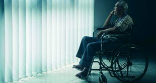
3. It is also called silent disease, as it occurs in the latter stage of life without any symptoms. However, it causes people to be bedridden with back pain, loss of height, kyphosis, pneumonia, and pulmonary thrombo-embolism. The treatment costs are high, after diabetes, hyperlipidemia, hypertension, and heart disease.
Diagnosis of osteoporosis
Diagnosis is made on the basis of bone mineral density level, which can be expressed in different ways:
• Absolute BMD (in the case of DXA gm/cm2).
• As a standard deviation (SD) score: it compares the individual’s result to a reference range, which should be for individuals of the same gender and ethnic background. Comparison may be to an age-matched population, in which case it may be referred to as a Z-score, or to a population of healthy young adults, in which case it is referred to as a T score.
• As a percentage: results are expressed in relation to an appropriate reference range.
• As a percentile: results are expressed in percentile that avoids the difficulty associated with percentage. This type of expressing result is not in practice.
BMD measurement techniques for osteoporosis
There are different modalities available for diagnosing osteoporosis, some of which are mentioned here:
RA (Radiographic Absorptiometry): In this method, the x-ray of hand and a small metal wedge are compared and the bone density of the middle phalanges is measured. Therefore, the measuring area is the hand.
Ultrasound method: It uses the sound of frequency above 20 KHz. Sound is transmitted faster in dense bone than osteopenic bone, and devices are calibrated against other methods to correlate with bone mass. Measuring area is the heel. It is suitable for younger perimenopausal women, but not for adult 50 years or above, as it underestimates the true prevalence of osteoporosis.
MRI (Magnetic Resonance Imaging): This method is considered an effective one, as it is non-invasive and radiation-free, and is reliable for assessing the features of the trabecular bone structure. Trabecular bone is highly responsive to metabolic stimuli and has a turnover rate approximately three to 10 times higher than cortical bone, and so it is a prime site for detecting early bone loss and monitoring response to therapeutic intervention. The spine, hip, or total body are measured in this method.
QCT (Quantitative Computed Tomography): The entire body is measured in this method. QCT refers to a class of technique in which the CT numbers, or x-ray attenuation, of a tissue is properly referenced to a calibration standard and then used to quantify some property of the tissue.
Laboratory tests: It measures the amount of collagen in urine samples that can indicate bone loss.
DXA (Dual Energy X-Ray Absorptiometry): DXA is considered as “gold standard” among clinical bone densitometry techniques. The precision of measurement by this technique is excellent (1%), and the radiation dose is very low. This method is best for women who are peri-menopausal (just becoming menopausal), and those who are newly menopausal. DXA is most commonly applied to scanning the lumbar spine, proximal femur, and whole body. X-rays of the spine, hip, and sometimes wrist are taken, and the bone density at each area is compared either with a reference population matched for age, gender, and ethnicity (Z score), or with a young and healthy individual matched for gender (T score). The results of these tests are compared with the results of same sex, healthy, young adults at the peak of their bone mass. This technology is new and expensive, and available in very few hospitals of Nepal. Therefore, limited studies are found by using DXA in Nepal.
Prevalence of osteoporosis
There is no homogeneity on the data of the prevalence of osteopenia and osteoporosis in the world. Different types of studies indicate the different prevalence of osteoporosis in the particular context only. The prevalence of osteoporosis is found to be very high in the world. Generally, one in two females and one in five males of age above 50 years suffer from osteoporotic fracture. There is no exact prevalence of osteoporosis in the context of Nepal. However there are few studies regarding osteoporosis from different perspectives. A study conducted among healthy people in the tertiary care health centers of Nepal by calcaneal ultrasonography reported the prevalence of osteoporosis and osteopenia at 22.4% and 60.6% respectively [1], while it was found to be 8.2 % in another study conducted in a tertiary hospital through the same technology [2]. In other studies conducted among adults of age 50 years and above in different hospitals of Kathmandu by using DXA, the prevalence of osteoporosis and osteopenia was 37.3 % and 38.5% respectively [3].
Risk factors of osteoporosis
The risk factors can be categorized into non-modifiable factors and modifiable factors.
Non-modifiable factors: Non-modifiable factors include gender, age, body size, ethnicity, heredity, genetics, etc.
Gender: Women have two to three times more chances of suffering from osteoporosis than men. It is because of the fact that women have two phases of age-related bone loss—a rapid phase initiated by a dramatic decline in estrogen produced by the ovaries that begins at menopause and last four-eight  years, followed by a slower continuous phase that lasts throughout the rest of life. The men go through only the slow, continuous phase. The slower continuous phase is caused by the combination of many factors, including age-related impairment of bone formation, decreased physical activity, and the loss of estrogen’s positive eff ect on calcium balance in the intestine and kidney, as well as its eff ects on bone. Age: Th e prevalence of osteoporosis increases with age. Th e prevalence of osteoporosis is 0 % in age 25- 34 years, 18.42 % in age 35-44 years, 18.18 % in 45-54 years, 21.42 % in age 55-64 years, and 50 % in age 65 years and above [4]. Body size: Small, thin women are more susceptible than larger women. Th e underweight have more probability of suff ering from osteoporosis. Body size and bone size are greater in obese people than in slender people, and peak bone mass is greater in obese people than in slender people. Ethnicity and heredity: Caucasian and Asian women are at greatest risk. African American and Hispanic women have a lower but significant risk. One study showed that Malay and Indian men had signifi cantly higher BMD than Chinese men at the lumbar spine [5]. Th ere were substantial race/ ethnic differences in BMD even within African or Asian origin [6]. However, no differences were found in the bone mineral density among diff erent ethnic groups in Nepal. It may be due to the fact that all Nepalis have almost same lifestyle and food consumption pattern. Genetics: If a parent suffers from osteoporosis or an osteoporotic bone fracture, the children tend to have reduced bone mass. Modifi able factors: It includes smoking, alcohol consumption, medication, low level of oestrogen in women, or low level of testosterone hormone. Cigarette smoking: Smoking decreases calcium absorption. It also reduces the production of oestrogen and leads to early menopause in women by causing the oestrogen to be metabolized quickly and reducing calcium absorption. Alcohol consumption: High intake of alcohol increases the amount of calcium lost in urine, which is associated with a reduction in bone mass, thus increasing susceptibility to the development of osteoporosis. Various studies show that moderate consumption of alcohol has positive effect on BMD, while long-term consumption of alcohol prevents formation of peak bone mass. Alcohol consumption disrupts the production of parathyroid hormone, which is a calcium regulating hormone.
Prevention of osteoporosis
Healthy life is our goal. This goal can be achieved with healthy bone. It consists of lifestyle behavior like good food habit, physical activity and avoiding smoking and alcohol consumption, etc.
Physical activity: Physical activity helps prevent fall-related fractures by improving muscle strength, body balance, and reaction time. It positively affects the bone density when calcium intake exceeds 1000 mg per day. It has been found that during extreme periods of inactivity, such as prolonged bed rest, bone loss occurs even when the level of calcium consumption is high. Resistance-training increases bone mass and prevents age-related declines in BMD.
Tea/coffee consumption: Different studies have found positive effect of tea consumption in osteoporosis. Tea and coffee contain caffeine that support the osteoblastic activities while suppressing the osteoclastic activities, hence reducing osteoporosis.
Dietary approach
Dietary behavior has a major role for the prevention of osteoporosis, as well as in the treatment. The dietary habit and food choice in osteoporotic patients was not suitable in Iran who had lower intake of zinc, calcium and vitamin D and higher intake of protein, iron and phosphorus than normal range [7]. The food habit, whether vegetarian or non-vegetarian, did not have any effect on osteoporosis [8]. However, as it contains high amount of calcium, 330 mg calcium in 100 gram fish [9], regular consumption of fish has prevented suffering from osteoporosis in Korea [10]. The RDA recommends a daily intake of 1000 mg to 1200 mg calcium to prevent osteoporosis and other diseases. The average intake of daily calcium consumption has been found to be 353.44 ±129.20 mg in pregnant women [11] and 520.44±296.97 mg in older people in Nepal, which is significantly low compared to the daily intake  requirement [3]. There are many calcium-rich foods in Nepal. Dairy products, fish, fruits, green vegetables, etc. can be consumed for the increment of calcium intake. Fresh water snail (ghongi in Tharu language) consumption is very common in the Terai region of Nepal, especially in the Tharu community. It contains very high amount of calcium (100 gram flesh of snail contains around 1321 mg calcium) while 100 ml milk contains only 120 mg calcium [9]. Most Nepalis depend on milk consumption for calcium intake. According to Commercial Agriculture for Smallholders and Agribusiness (CASA) report 2020, the requirement for milk in Nepal is 92 liters per person, and the country produces 72 liters per person annually, thus fulfilling 80% of this requirement. Calcium supplementation as well as fortified food is also preferred to the risk groups such as older people, pregnant women, lactating mothers, diseased people, etc.
Another dietary intake for osteoporosis is vitamin D, which can be achieved from food only up to 10 to 15%, especially from animal source. It can be mainly fulfilled by sun exposure (up to 85 %). Therefore, vitamin D is also called sunshine vitamin. Around 10 to 15 minutes sun exposure to the naked body is sufficient for the formation of vitamin D in our body. There are few countries where there is no sun ray available during the year. Vitamin D supplementation is required in that context. In Nepal, thankfully, there are no large buildings and infrastructures, so exposure to the sun is not a big issue.
However, the lifestyle of urban people is changing. Food habit is also changing. Consequently, more than two third (73.6%) of people suffer from vitamin D deficiency in Kathmandu, of which 21.08 % are male and 52.61% are female [12]. It is a serious matter. Physiologically, vitamin D is the precursor for absorption of calcium in our body. Without sufficient vitamin D, no absorption of calcium occurs in our body.
Another nutrient concerned with osteoporosis is vitamin C, which enhances absorption of calcium. When taken together, they can maximize bone strength and may play a role in preventing osteoporosis. Foods having plenty of vitamin C are citrus fruits like orange, amala, lemon, and leafy green vegetables, etc. Besides these, other nutrients like protein, magnesium, vitamin K, zinc, etc. should be consumed in sufficient amounts for strong bone.
The nutrient that we have to limit for osteoporosis is excess salt, because it can cause our body to release calcium, which is harmful to the bones. Recommended Dietary Allowance (RDA) recommends avoiding foods that contain more than 20% of the daily recommended value for sodium. We have to limit its intake, no more than 2,300 mg per day whenever possible. For our daily household measurement, one tablespoon contains 10 g salt, which contains 3,900 mg sodium. Therefore, salt intake should be minimized to 2 g per day. Processed food like smoked, cured, salted, or canned meat, fish or poultry, frozen breaded meats, etc. contains high amounts of salt.
Conclusion
The concept of nutrition and dietetics is new in Nepal. It is mostly considered in the case of renal, cardiovascular, and diabetes disease, but less prioritized in other disciplines. However, it is multidisciplinary. As osteoporosis is inter-related to orthopedics, obstetrics and gynecology, nephrology, endocrinology, etc., and all stages of life are prone to osteoporosis, it has to be considered for further evaluation. In addition to these, the least number of people, even few medical professionals, know about osteoporosis. Therefore, proper policy and strategy should be made for the prevention of osteoporosis so that a healthy society could be developed in the coming days.
References:
1. Bagudai S, Upadhayay HP. Prevalence of Osteoporosis and Osteopenia Status among Nepalese Population using Calcaneal Ultrasonography Method. J Coll Med Sci. 2019;15: 249–255. doi:10.3126/jcmsn.v15i4.24008
2. Shrestha S, Dahal S, Bhandari P, Bajracharya S, Marasini A. Prevalence of osteoporosis among adults in a tertiary care hospital: A descriptive cross-sectional study. J Nepal Med Assoc. 2019;57: 396–400. doi:10.31729/jnma.4753
3. Chaudhary NK, Timilsena MN, Sunuwar DR, Pradhan PMS, Sangroula RK. Association of Lifestyle and Food Consumption with Bone Mineral Density among People Aged 50 Years and above Attending the Hospitals of Kathmandu, Nepal. J Osteoporos. 2019;2019. doi:10.1155/2019/1536394
4. Sharma S, Tandon VR, Mahajan A, Kour A, Kumar D. Preliminary screening of osteoporosis and osteopenia in urban women from Jammu using calcaneal QUS. Indian J Med Sci. 2006;60: 183–189. doi:10.4103/0019-5359.25679
5. Yang PLS, Lu Y, Khoo CM, Leow MKS, Khoo EYH, Teo A, et al. Associations between ethnicity, body composition, and bone mineral density in a southeast Asian population. J Clin Endocrinol Metab. 2013;98: 4516–4523. doi:10.1210/jc.2013-2454
6. Nam HS, Shin MH, Zmuda JM, Leung PC, Barrett-Connor E, Orwoll ES, et al. Race/ethnic differences in bone mineral densities in older men. Osteoporos Int. 2010;21: 2115–2123. doi:10.1007/s00198-010-1188-3
7. Mahdaviroshan M, Ebrahimimameghani M. Assessments of dietary pattern and nutritional intake in osteoporotic patients in Tabriz. J Paramed Sci. 2014;5: 27–30. doi:10.22037/jps.v5i3.6369
8. Ho-Pham LT, Nguyen PLT, Le TTT, Doan TAT, Tran NT, Le TA, et al. Veganism, bone mineral density, and body composition: A study in Buddhist nuns. Osteoporos Int. 2009;20: 2087–2093. doi:10.1007/s00198-009-0916-z
9. Nepal Government. Food Composition Table For Nepal. 2012.
10. Choi E, Park Y. The association between the consumption of fish/shellfish and the risk of osteoporosis in men and postmenopausalwomen aged 50 years or older. Nutrients. 2016;8. doi:10.3390/nu8030113
11. Acharya O, Zotor FB, Chaudhary P, Deepak K, Amuna P, Ellahi B. Maternal Nutritional Status, Food Intake and Pregnancy Weight Gain in Nepal. J Health Manag. 2016;18: 1–12. doi:10.1177/0972063415625537
12. Rai CK, Shrestha B, Sapkota J, Das JK. Prevalence of vitamin D deficiency among adult patients in a tertiary care hospital. J Nepal Med Assoc. 2019;57: 226–228. doi:10.31729/jnma.4534than
 Medicosnext


